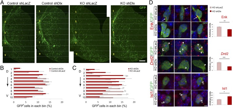
Fig. 6.
Knocking down Dlx1/2 genes alleviates abnormal patterns of cell migration and differentiation in the Nolz-1 KO striatum. (A) shDlx1/2 plasmids are coelectroporated with GFP reporter plasmids into the E13.5 LGE by in utero electroporation. The migratory pattern of shDlx1/2-GFP+ cells is analyzed at E18.5. In control brains, the distribution pattern of shDlx1/2-GFP+ cells is similar to that of mock control shLacZ-GFP+ cells. In Nolz-1 KO brains, the number of shDlx1/2-GFP+ cells is decreased in the ventral striatum compared to that of shLacZ-GFP+ cells. (B and C) The striatum is divided into 10 bins along the dorsoventral axis. The percentage of shDlx1/2-GFP+ cells in each bin does not differ from that of shLacZ-GFP+ cells in control brains (B). In Nolz-1 KO brains (C), the percentage of shDlx1/2-GFP+ cells is decreased in the ventral striatum (bins nos. 7–9) but is concurrently increased in the dorsal striatum (bin no. 3) compared to that of shLacZ-GFP+ cells. (D) Electroporation of shDlx1/2 plasmids into the E13.5 Nolz-1 KO LGE decreases Enk (arrows) and Drd2 mRNAs (arrowheads) but increases Isl1 mRNA expression (double arrows) in the E18.5 KO striatum compared to the mock control of shLacZ. D, dorsal; V, ventral. *P < 0.05; ***P < 0.001; n = 3/group. (Scale bars, 100 μm [A] and 5 μm [D].)

