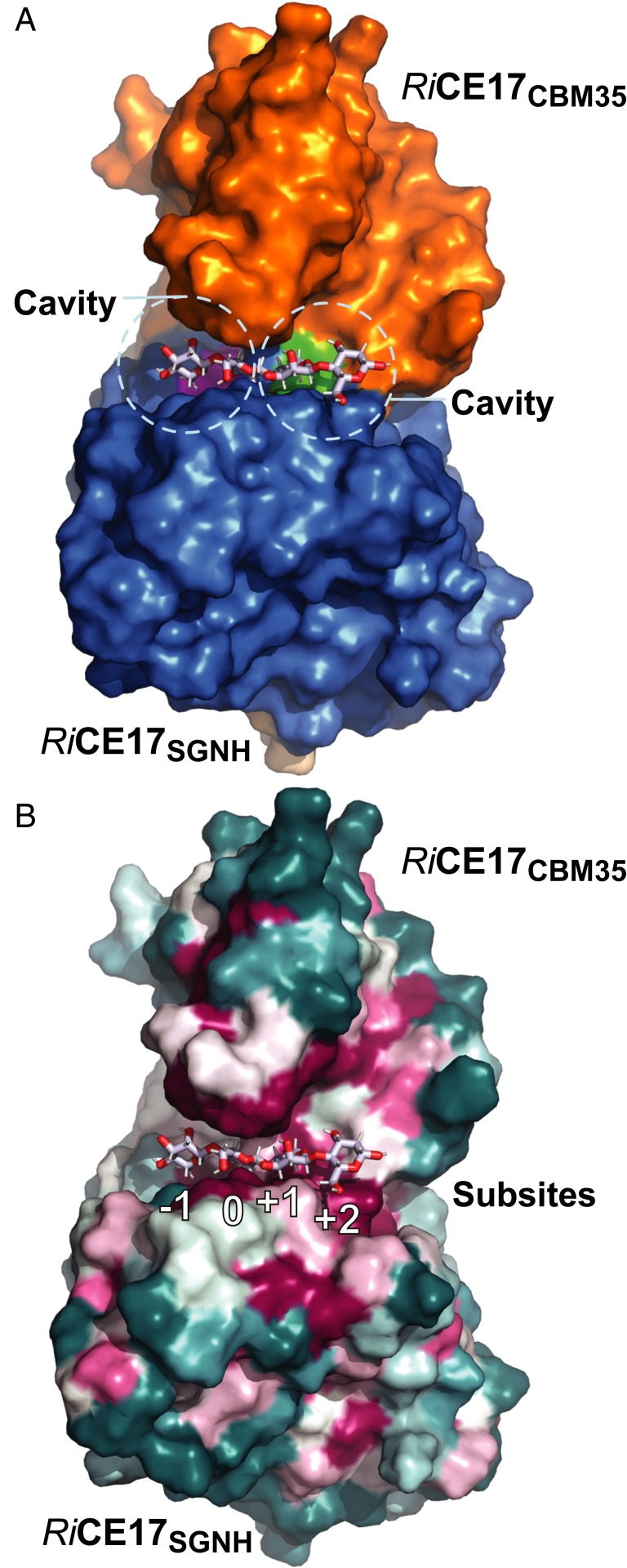
Fig. 3.
The 3D structures of substrate and product complexes of RiCE17. (A) The magenta and green patches in the interface between the RiCE17 catalytic domain and the RiCBM35 domain indicate cavities on the enzyme surface that may bind galactose residues decorating galactoglucomannan at the C6 position. In B the conservation score derived by ConSurf is projected on the RiCE17 surface (highly conserved amino acid residues [magenta], semiconserved [white], and variable residues [blue] are indicated). The highly conserved residues are concentrated around the substrate-binding site. Subsites are labeled below the substrate according to the nomenclature suggested by Hekmat et al. (37).

