Fig. 3.
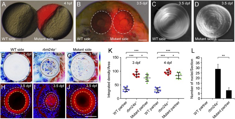
Rescue of lens fiber cell denucleation but not lens transparency in rbm24a mutants by heterotypic parabiosis. (A) Parabiosis by surgically joining an unlabeled WT embryo and a rhodamine-lysine dextran-labeled rbm24a mutant (red). (Scale bar, 200 µm.) (B) Parabiotic WT and rbm24a mutant partners at 3.5 dpf, with eyes outlined by dashed circles. (Scale bar, 100 µm.) (C and D) Differential interference contrast images show the lens phenotypes in parabiotic WT and mutant partners. (Scale bar, 50 µm.) (E–G) Lens transparency determined by confocal reflection mode, with vertical color bar indicating colors of 10 focal planes at a depth of 30 µm. (Scale bar, 50 µm.) (H–J) Staining of lens sections with phalloidin and Hoechst 33342 shows lens fiber cell denucleation in WT, rbm24a mutants, and rbm24a mutants parabiosed with WT embryos. (Scale bar, 50 µm.) (K) Scatterplot comparing lens transparency by analyzing the integrated density of the scanned lens region in the maximum projection image obtained under the confocal reflection mode. Horizontal lines and error bars represent the mean value ± SD from six to nine independent lenses (*P < 0.05; ***P < 0.001, Student’s t test). (L) Statistical analysis of lens fiber cell denucleation by examining the number of fiber cell nuclei present within 2/3 radius of a lens section at the level of the optic nerve (outlined by dashed yellow circles in H–J). This area shows complete denucleation in WT embryos at 3.5 dpf. The results were obtained from three independent lens sections (**P < 0.01, Student’s t test).

