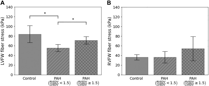
Fig. 8.
Comparison of peak myofiber wall stress between control and pulmonary arterial hypertension (PAH) groups in the left ventricular free wall (LVFW; A) and right ventricular free wall (RVFW; B) regions. RVEDV/LVEDV, ratio of right ventricular end-diastolic volume to left ventricular end-diastolic volume. *P < 0.01, statistical significance between groups.

