
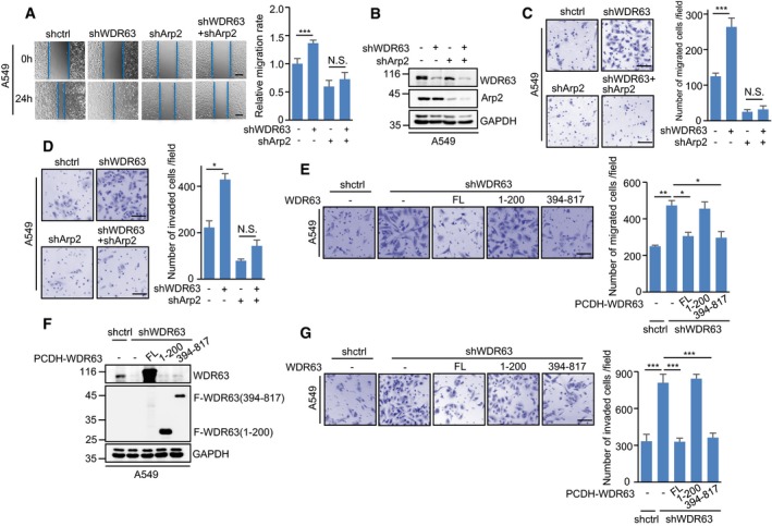
Figure 5. WDR63 inhibits cell migration and invasion through the Arp2/3 complex.
-
A–DA549 cells expressing control shRNA, WDR63 shRNA#1, Arp2 shRNA, or both WDR63 and Arp2 shRNA were subjected to wound‐healing (A), transwell migration (C), and transwell invasion (D) assays. Lysates from these cells were also analyzed by Western blot (B). The shown images are representative of three independent experiments. Data shown are mean ± SD (n = 3). *P < 0.05; ***P < 0.001; N.S., no significance; one‐way ANOVA. Scale bar in (A): 200 μm. Scale bars in (C and D): 100 μm.
-
E–GA549 cells expressing either control shRNA or WDR63 shRNA#1 were infected with lentiviruses expressing Flag‐tagged full‐length or truncated WDR63 proteins as indicated. Cells were then subjected to transwell migration (E) and transwell invasion (G) assays. Lysates from these cells were also analyzed by Western blot (F). The shown images are representative of three independent experiments. Data shown are mean ± SD (n = 3). *P < 0.05; **P < 0.01; ***P < 0.001; one‐way ANOVA. Scale bar: 100 μm.