
Figure EV2. Telomere fusion assay by PCR.
-
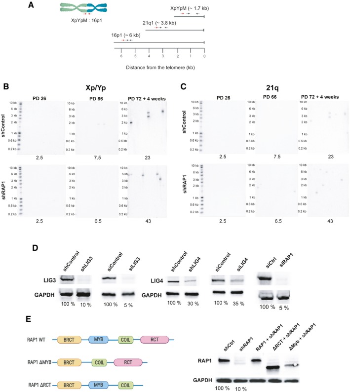
ALocation of the primers and probes used for the telomere fusion PCR: Red arrows indicate positions of subtelomeric PCR primers, while black arrows represent the primers used to generate DNA probes for radioactive hybridization with the Southern blot membranes.
-
B, CRepresentative Southern blot membranes of the telomere fusion PCR assay shown in Fig 2A and B. Membranes were subsequently hybridized with the 16p probe, XpYp probe, and finally with the 21q probe.
-
DDifferent RAP1 constructs used in the telomere fusion assay of senescent MRC‐5 cells. The percentage of protein expression is indicated below the blots.
-
EExpression of RAP1, LIG3, and LIG4 related to Fig 2C. The percentage of protein expression is indicated.