
Figure EV1. Characterization of Mex3a expression pattern in murine tissues.
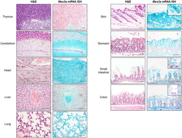
H&E staining and Mex3a mRNA ISH in serial sections of different mouse organs at postnatal day 17. Each punctuate red dot in the ISH panels represents a hybridization event with a single Mex3a mRNA molecule. Inserts depict high magnification of the boxed areas. The diffuse signals observed in the liver are the result of non‐specific staining. Scale bars, 50 μm.