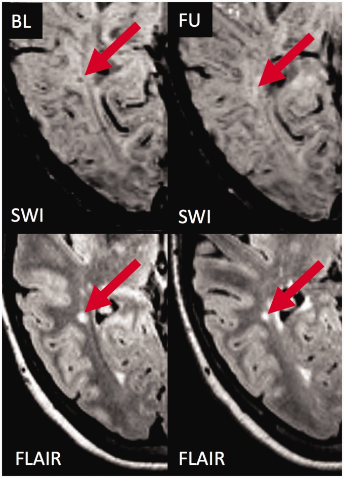
Figure 2.
The fate of an exemplary ‘core sign’ lesion without contrast enhancement.
Susceptibility weighted imaging (SWI; top) and T2 weighted fluid attenuated inversion recovery (T2w-FLAIR; bottom) images are shown at baseline (left) and follow-up (right). At baseline, a hyperintense white matter lesion is visualised on T2w-FLAIR. The lesion is characterised by a SWI hypointense signal within the centre of the lesion (‘core sign’). During follow-up, the lesion became smaller on T2w-FLAIR, and the SWI hypointense signal (‘core sign’) disappeared rendering the lesion isointense to slightly hyperintense on SWI. BL: baseline; FU: follow-up.

