
Figure 1.
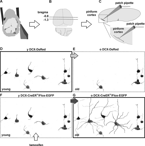
Experimental model. (A–C) Acute brain slice preparation and position of the piriform cortex. After dissection (A), the brain of adult mice was dissected and acute coronal slices were collected from (B) the rostral part of the posterior piriform cortex (Bregma = −0.8 to −1.3), where tangled and complex cells are most abundant. In acute slices, tangled and complex cells were scattered unevenly within the piriform cortex. (D–E) Schematic representation of fluorescently labeled cells in the adult brain of DCX-DsRed mice. In the piriform cortex of 2- to 4-month-old (y) DCX-DsRed mice, tangled cells and immature young complex cells are fluorescently labeled (D). In the 4- to 8-month-old (o) DCX-DsRed mice, the number of fluorescently labeled cells decreases dramatically, as DCX is no longer expressed (E). (F, G) Schematic representation of fluorescent-labeled cells in the adult brain of DCX-CreERT2/Flox-EGFP mice, after tamoxifen treatment. In young DCX-CreERT2/Flox-EGFP mice, immature tangled and complex cells are labeled fluorescently (F). In old DCX-CreERT2/Flox-EGFP mice, cells that expressed DCX at the moment of tamoxifen treatment are permanently labeled fluorescently. Thus, targeting labeled cells in young DCX-DsRed mice (D, bold frame) allows characterization of the more immature tangled and complex cells. Targeting labeled cells in old DCX-CreERT2/Flox-EGFP mice (G, bold frame) allows characterization of the more mature old complex cells. Note: for illustrative purposes, the number of fluorescent cells has been enhanced in regard to their actual density in the tissue (see Rotheneichner et al. 2018).