
Figure 2.
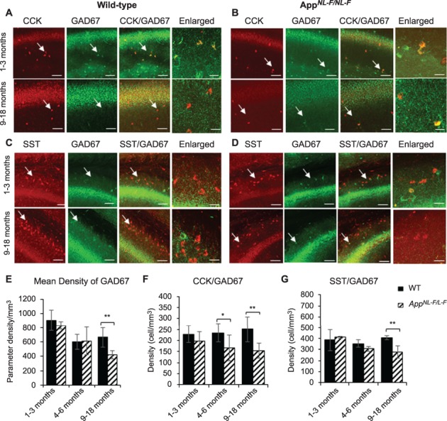
CCK- and SST-expressing interneurons show an age-dependent loss in the AppNL-F/NL-F AD model. (A–D) Confocal microscope Z-stack images showing the expression of CCK- and SST-expressing cells (both in red channel), colocalized with GAD67, the marker for GABA production (green channel) in 1–3- and 9–18-month-old wild-type age-matched AppNL-F/NL-F mice in CA1. Images taken at ×20 magnification (scale bar = 50 μm) and ×63 magnification (enlarged images, scale bar = 20 μm). In aged AppNL-F/NL-F mice, CCK- and SST-positive cells were found to be weakly colocalized with GAD67 compared with the 1–3-month-old AppNL-F/NL-F mice. (E–G) The graphs represent mean density of GAD67-, CCK-, and SST-positive cells in wild-type age-matched AppNL-F/NL-F measured at three ages: 1–3 months, 4–6 months, and 9–18 months. Overall, GAD67, CCK, and SST expression showed an age-dependent decline in the AD model, which was significantly different from their control wild-type counterparts at 9–18 months. However, CCK cells also showed a significant decline in AppNL-F/NL-F mice 4–6. A two-way ANOVA was performed with pairwise comparisons corrected for multiple comparisons (α = 0.05), with a post hoc Tukey’s test for multiple comparisons. *P < 0.05, **P < 0.001.