
Figure 2.
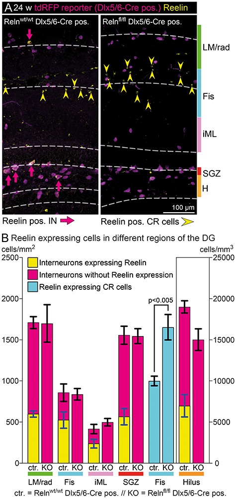
Reelin expression pattern in the mature dentate gyrus. (A) Immunohistochemical staining for tdRFP and Reelin was used to analyze the total number of INs, the number of Reelin-expressing INs and Reelin-expressing CR cells in different areas of the dentate gyrus. INs were identified by tdRFP reporter staining, while CR cells were identified as small Reelin-expressing cells without tdRFP expression. (B) Quantification of cell numbers in different layers. The total number of INs and their distribution within the dentate gyrus was unchanged in IN-specific Reelin knockout mice. In addition, as expected, all INs proved to be Reelin-negative in this model. Interestingly, the number of Reelin-expressing CR cells was significantly increased in adult IN-specific Reelin knockout mice (t-test; N = 5; age: 24–30 weeks; error: ±SEM), and the number of additional CR cells (654 ± 108) corresponded well to the number of INs that lost Reelin expression within a distance of 60 μm around the fissure (Fis) (524 ± 99). Abbreviations: LM/rad—Stratum lacunosum-moleculare and parts of stratum radiatum; Fis, fissure (±60 μm around the hippocampal fissure); iML, inner molecular layer; SGZ, subgranular zone (±10 μm around the inner border of the granule cell layer); and H, hilus.