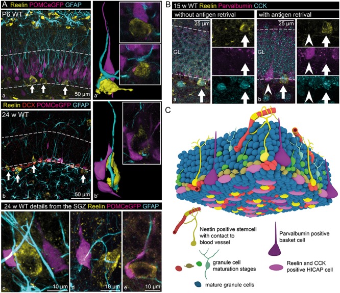
Figure 7.

Reelin-expressing inhibitory INs within the stem cell niche. (A) Reelin-expressing INs (white arrows) could be found within the SGZ of the dentate gyrus of young (P6 – a, a’) and adult mice (24 weeks – b, b’). Immunohistochemical staining for Reelin, glial fibrillary acidic protein (GFAP, labeling glial cells including RGLCs, Doublecortin (DCX), and POMCeGFP (Overstreet et al. 2004) (both labeling immature neurons) revealed close contact between Reelin-expressing INs, RGLCs, and immature neurons (Images a–e and 3D-reconstructions a’, b’). (B) Immunohistochemical stainings of adult (15 weeks) wild-type mice for Reelin, Parvalbumin (marker for basket cells) and CCK (marker for HICAP cells) in the SGZ (staining performed without (a) and with acidic antigen retrieval (b)). The staining revealed coexpression of Reelin and CCK in INs (full arrows), while the Parvalbumin-positive cells were never immunoreactive for Reelin (arrowheads). (C) A representative scheme depicts the observed integration of Reelin-expressing INs within the subgranular stem cell niche (general morphology of the stem cell niche based on data by Fuentealba et al. (2012) and Kempermann et al. (2015)).
