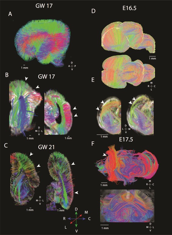
Figure 2.

Diffusion MR tractography of cortices at GW 17–21 (A–C) and mice at E 16.5–E 17.6 (E, F) shows major differences in the organization of fibers spanning the cortex. In humans, many fibers course radially within the developing cortex. At GW 17 whole brain tractography (A) and 1 mm thick coronal slices through the anterior and posterior regions of the cortex (B) show fibers coursing from the proliferative zones to the CPs (white arrowheads). A similar situation is observed at GW 21 (C) where coronal slices set to capture fibers coursing through these slices identify radial fibers spanning the proliferative pool and the CP in the anterior and posterior cortex (white arrowheads). In contrast, diffusion MR scans of mice at E 16.5 (D, E) and at E 17.5 (F) show fibers principally organized across the medial to lateral axis (white arrowheads). (D) Whole brain tractography of mice show fibers coursing across the medial to lateral and dorsal to ventral direction through the developing cortex. (E) Coronal slices through the rostral and caudal cortex at E 16.5 capture fibers coursing tangential to the cortical surface rather than radially (white arrowheads). (F) At E 17.5, a similar situation is observed in the mouse where fibers are observed coursing across the medial to lateral direction (white arrowheads). Humans at GW 17–21 and mice at E 16.5–17.5 were selected for comparison because they are at roughly equivalent ages of development (Workman et al. 2013). The following abbreviations are used: A: anterior; P: posterior; R: rostral; C: caudal; M: medial, L: lateral; D: dorsal; and V: ventral.
