
Figure 3.
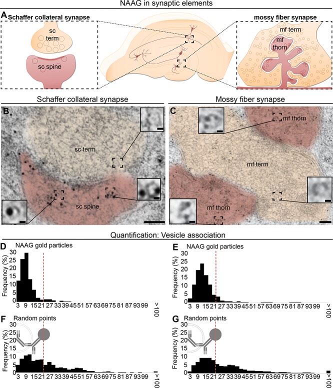
NAAG is located in vesicular structures in both postsynaptic dendritic spines in stratum radiatum (A, left) and in postsynaptic mossy fiber thorns in dentate gyrus (A, right). In the dendritic spines and thorns, gold particles representing NAAG were closely related to vesicular structures, which in size and shape resemble presynaptic vesicles (insets in B and C). Scale bars in (B and C): 100 nm; scale bar in insets: 10 nm. The distribution of distances of gold particles and the closest vesicle-like structures was quantified. In both postsynaptic compartments, the sc spines and the mf thorns, a large proportion of the NAAG particles were localized within 21 nm from the center of a vesicle (D and F). For comparison, for the randomly inserted points (E and G), only about half of the points were found within the same range from the vesicle (P = 9.1 × 10−43 for sc spines; and P = 8.9 × 10−50 for mf thorns; Pearson chi square test). 21 nm represents the theoretical distance between the center of the gold particle and the epitope (the radius of the gold particle [5 nm] + the length of the primary–secondary complex).