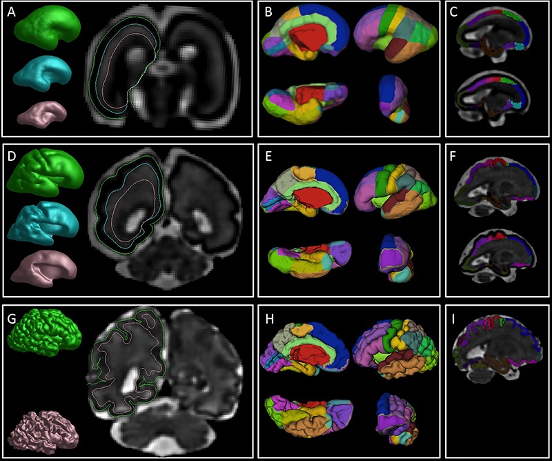
Figure 2.

An overview of the image processing steps. First column: reconstruction of the pial (green), SP (cyan), and IZ (pink) surfaces in 25 (A), 29.57 (D), and 36.86 (G) GW brain. Note the borders of the reconstructed surfaces that are superimposed on the coronal sections and mark the border between fetal compartments. Second column: manually painted cortical ROIs superimposed on the reconstructed pial surface in 25 (B), 29.57 (E), and 36.86 (H) GW brain. Third column: volumetric ROIs of CP (upper row in C, F, and I) and SP (bottom row in C and F) in 25 (C), 29.57 (F), and 36.86 (I) GW brain.
