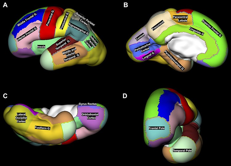
Figure 3.

An example of 24 ROIs manually segmented on the reconstructed CP surface (lateral (A), medial (B), ventral (C), and anterior (D) views) of the left hemisphere in 29.57 GW fetus. Note that the middle and inferior temporal gyri were merged with temporal pole and fusiform gyrus into one region called latero-inferior temporal neocortex.
