Figure 1.

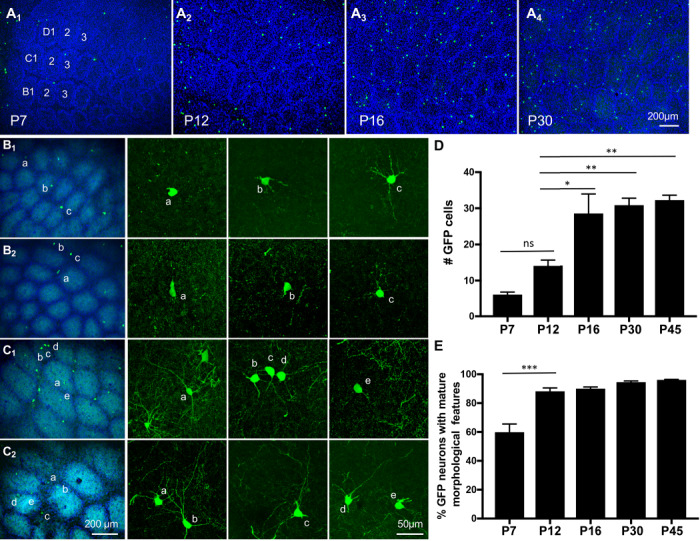
GAD67-GFP/GIN-expressing neurons arise and mature in the somatosensory barrel cortex at late postnatal stages. (A) Images of GFP-expressing neurons in tangential sections of the PMBSF at indicated ages. Barrels in arcs 1–3 of rows B-D are indicated in the image of P7 PMBSF. Noticing preferential localization of GFP+ cell bodies (green) in barrel walls outlined by densely packed DAPI-labeled nuclei (blue). Timing of GFP+ cells arising in the entire neocortical sheet is presented in Supplementary Fig. S2. (B-C) Representative images of GFP+ cell morphology at P7 (B1, B2) and P12 (C1, C2). The first column from the left shows low-magnification images of GFP+ neurons (green) in the PMBSF overlay with Vglut2-immunolabeled TCA (cyan) and DAPI counterstain of cell nuclei (blue), and neurons marked by letters are shown in confocal images in corresponding right panels. Neurons with simple morphology as displayed by neurons a in B1,a, b in B2, and e in C1 are considered immature neurons, while longer and more complex neurites shown by the other neurons are considered as mature morphological features. (D-E) Quantification of the number of GFP+ cells in nine barrels in arcs 1–3 of rows B-D in PMBSF (D), and the fraction of the neurons displaying mature morphological features at indicated ages (E). GFP+ neurons in serial tangential sections through the PMBSF were analyzed, and data represent mean per 50 μm section ± SEM, N ≥ 3 mice for each age group. *P < 0.05, **P < 0.01, ***P < 0.001, and one-way ANOVA followed by Tukey post hoc tests.
