Figure 3.

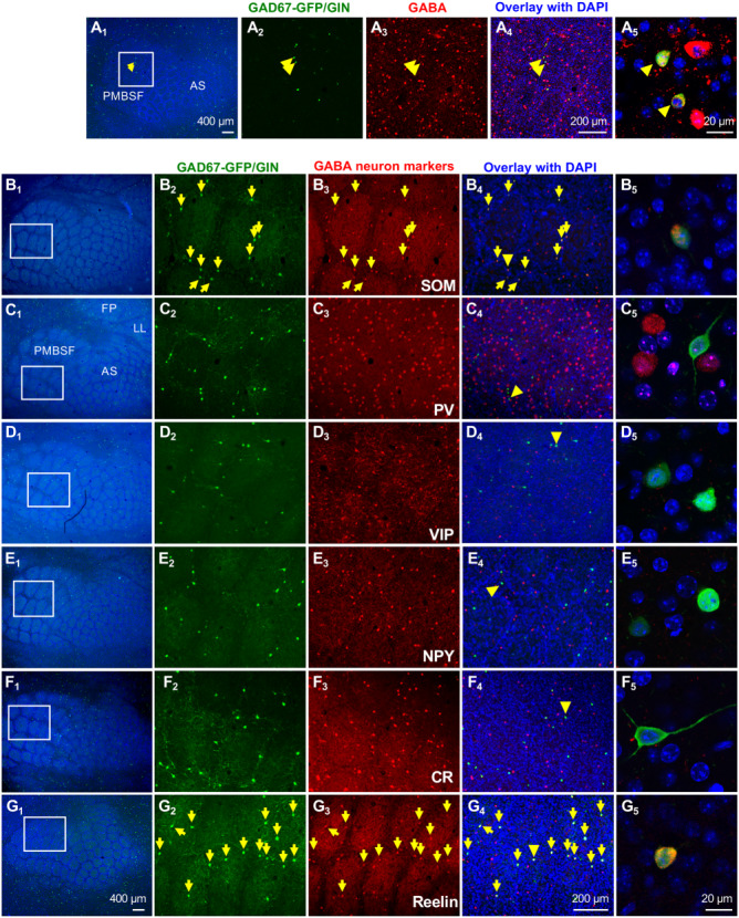
Barrel Cortex GAD67-GFP/GIN-expressing neurons represent a population of Reelin- and SOM-expressing GABAergic neurons. All images shown are tangential sections of the barrel cortex from P30 mice. (A) GFP and GABA double immunostaining. A2–A4 show GFP (green) and GABA (red) staining with DAPI counterstain (blue) in the PMBSF area outlined by a white box in A1.A5 shows a higher magnification confocal image of colocalization of GFP and GABA immunoreactivity in two neurons indicated by arrowheads. Noticing that GABA+ cell bodies display a ring patterning (barrel walls), and GFP+ neurons are preferentially localized to the ring. (B-G) Immunostaining for GFP and GABAergic neuron subtype markers. B1–G1 show low-magnification images of the barrel cortex with TCA immunolabeled by Vglut2 (cyan)and DAPI counterstain of the cell nuclei (blue). Receptive fields corresponding to specific body parts, whiskers (PMBSF), anterior snouts (AS), lower lip (LL), and forepaw (FP) are indicated in C1. Higher magnification images of staining of GFP, indicated markers and overlay with DAPI in the regions outlined by white boxes are shown in B2, B3, and B4–G2, G3, and G4, respectively. Neurons stained by both GFP and indicated markers are pointed by yellow arrows. Confocal images of GFP+ neurons indicated by yellow arrowheads in B4–G4 are shown in B5–G5, and single-channel confocal images for individual staining of these neurons are presented in Supplementary Fig. S3. SOM and Reelin immunoreactivities were detectable in nearly all GFP+ cell bodies in the barrel cortex. No GFP+ neuron showed immunoreactivity for PV or VIP. Rarely, GFP+ neurons showed colocalization with NPY or CR (Supplementary Fig. S4). Quantification of colocalization of GFP with each marker is presented in Table 1.
