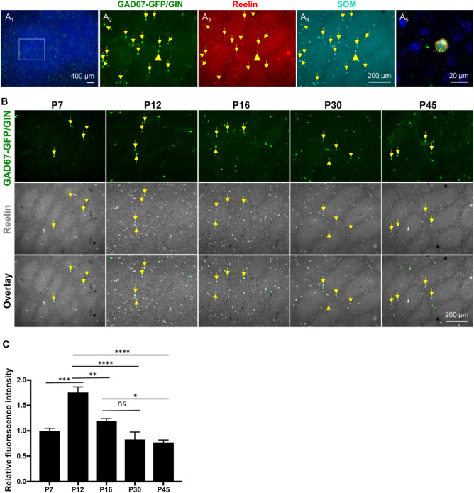
Figure 4.

Reelin immunoreactivity in GAD67-GFP/GIN-expressing neurons correlates with their morphological maturation in postnatal barrel cortex. (A) GFP, Reelin, and SOM triple immunostaining show GAD67-GFP/GIN-expressing neurons as a subpopulation of SOM+ neurons expressing Reelin (yellow arrows) in the barrel cortex at P30. (A1) A low-magnification image with DAPI counterstain of the cell nuclei (blue). (A2—A4) Higher magnification images of GFP (green), Reelin (red) and SOM (cyan) staining in the region outlined by a white box in A1. (A5) A representative confocal image of colocalization of both Reelin and SOM in a GFP+ neuron indicated by yellow arrowheads in A2—A4. (B-C) Developmental profiling of Reelin immunoreactivity in GAD67-GFP/GIN-expressing neurons in the barrel cortex. (B) Representative images of double immunostaining of GFP (green) and Reelin (gray) of the PMBSF at indicated ages. (C) Fluorescence intensity of Reelin immunostaining in GFP+ neurons at indicated ages normalized to that at P7, indicating a peak of Reelin immunoreactivity in GFP+ neurons at P12. Data represent mean ± SEM, N = 3–4 mice per time point, with 10–15 neurons analyzed per mouse. *P < 0.05, **P < 0.01, ***P < 0.001, ****P < 0.0001, and one-way ANOVA followed by Tukey post hoc tests.
