Figure 5.

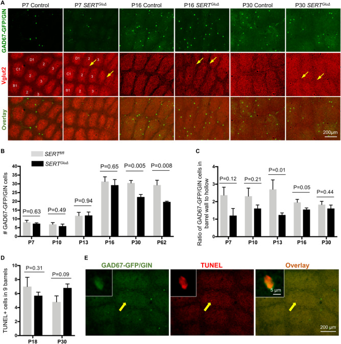
The number and spatial distribution of GAD67-GFP/GIN-expressing neurons in the barrel cortex of SERTGluΔ mice. (A) Representative images of GFP-expressing neurons in tangential sections of the PMBSF from SERTGluΔ and control littermate mice at indicated ages. Barrels in arcs 1–3 of rows B-D are indicated in the P7 cortex. Boundaries between barrels are blurred (arrows) in SERTGluΔ mice at P7 as well as at P16 and P30, while the gross barrel architecture is preserved. (B-C) Quantification of the number of GFP+ neurons and their distribution in nine barrels of arcs 1–3 of rows B-D of the PMBSF. (B) Significant reduction in the number of GFP+ neurons in one- and two-month-old SERTGluΔ mice as compared to control littermates. The numbers of GFP+ neurons at earlier developmental stages were comparable between the two genotypes. (C) Evaluation of the spatial distribution of GFP+ neurons in the barrels, by quantifying the ratio of GFP+ neurons in the barrel walls relative to that in the barrel hollows in SERTGluΔ and control mice. For each time point, serial sections through the PMBSF from SERTGluΔ and control littermates were processed and analyzed in parallel. Data represent mean per 50 μm section ± SEM, N = 4–6 mice per age per genotype per time point, and P values are based on t-tests. (D-E) Assessing apoptosis by TUNEL assay. (D) Quantifying the total number of TUNEL+ cells in arcs 1–3 of rows B-D in serial tangential sections through the PMBSF. N = 5 mice for each genotype per time point. Data represent mean ± SEM, t-tests. (E) Representative images showing TUNEL in a GFP+ neuron located in the barrel cortex at P30. Left corner insets show confocal images of the labeled cell indicated by a yellow arrow. The TUNEL+ cell body was smaller, compared to other GFP+ cell bodies in the same brain section, presumably due to shrinking of the dying cell.
