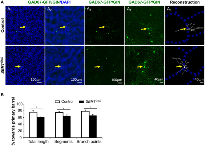
Figure 6.

Disrupting SERT expression in developing TCA alters GAD67-GFP/GIN-expressing neuron dendrite patterning in the cortex. (A) Examples of reconstruction of the dendritic tree of GFP+ neurons located close to the edge of barrel walls in the PMBSF of P30 SERTGluΔ and control littermate mice. (A1) Low-magnification images showing GFP+ cell bodies (green) at barrel walls delineated by a high density of DAPI-stained cell nuclei (blue) on tangential sections of the PMBSF. (A2) Original images processed using the mean filter and bandpass filter tools of ImageJ software to define barrel walls. (A3) GFP+ cells pointed by yellow arrows chosen for reconstruction. (A4) Higher magnification confocal images of GFP+ dendritic profiles. (A5) Reconstruction of GFP+ dendritic trees superimposed over barrel walls. (B) Quantification of the distribution of the dendrite length, dendritic segments and branching points of GFP+ neurons located at barrel walls relative to corresponding primary barrels in the PMBSF of P30 mice. Each bar represents mean ± SEM, N = 3 mice per genotype, with five neurons analyzed for each SERTGluΔ mouse and 4–6 neurons analyzed for each control mouse, *P < 0.05, t-tests.
