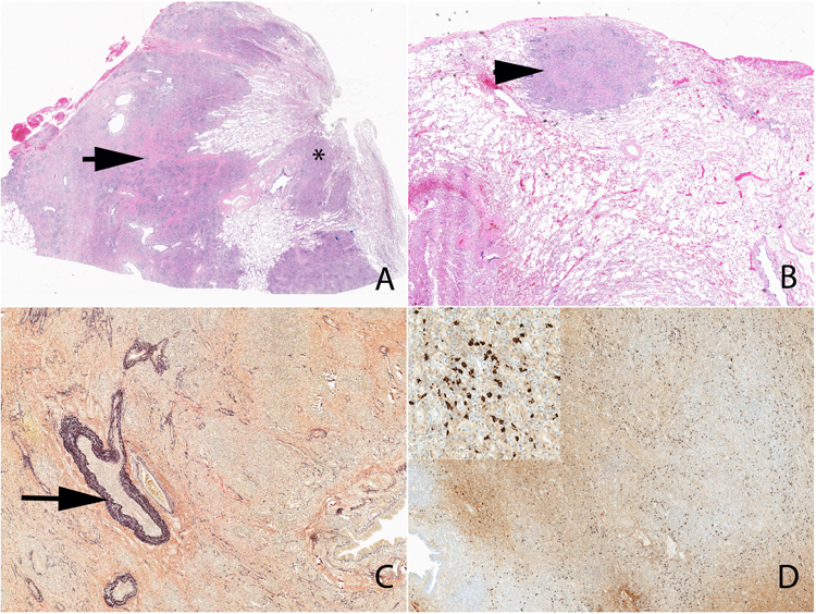
Figure 10.

Low power view of IgG4-related pulmonary disease. Note the tumefactive lesion (arrow) (A), extension along the bronchovascular tree (A) (*) and subpleural involvement (B) (arrowhead). An elastic stain highlights the focus of obliterative phlebitis (C) (arrow). An immunohistochemical stain shows a diffuse increase in IgG4+ plasma cells (D and inset).
