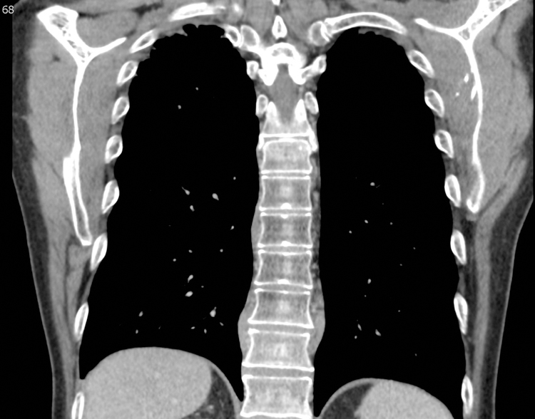
Figure 3.

Bilateral paravertebral masses in a 61 year-old-male with IgG4-RD. Coronal reformatted image from a Chest CT scan at the level of the thoracic spine on soft tissue windows. There are bilateral paravertebral masses, seen on the right at T8 and T10/11 and on the left at T10/11 (arrows). Biopsy confirmed IgG4-RD.
