Figure 4.

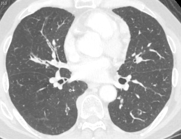
Bronchial wall thickening in 61 year-old-male with cough and IgG4-RD. Axial Chest CT scan on lung windows demonstrates diffuse, multifocal nodular thickening of the bronchial walls, best seen in the right middle lobe (arrow).

Bronchial wall thickening in 61 year-old-male with cough and IgG4-RD. Axial Chest CT scan on lung windows demonstrates diffuse, multifocal nodular thickening of the bronchial walls, best seen in the right middle lobe (arrow).