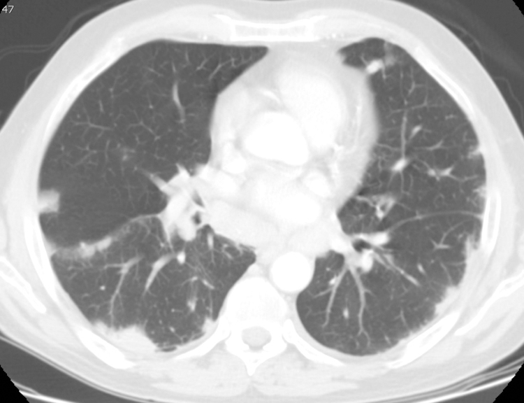
Figure 6.

Peripheral nodules and consolidation in 69-year-old male with IgG4-RD. Chest CT axial scan at the level of the aortic root on lung windows. There are multiple, bilateral peripheral lung nodules and consolidative opacities, that also extend along the right major and minor fissures, in a perilymphatic distribution that is characteristic of IgG4-RD. The patient denied pulmonary symptoms.
