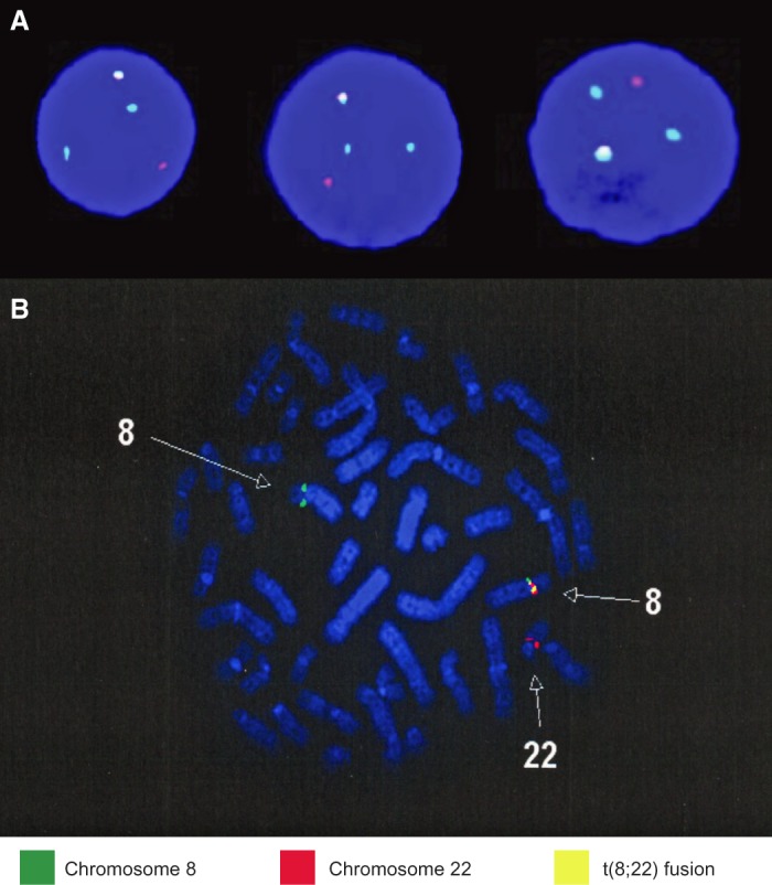
Figure 2.

Fluorescence in situ hybridization (FISH) panel. The FISH panel results identify the presence of the t(8;22) translocation in both patients. Two hundred cells were analyzed for disruption in FGFR1 using FGFR1 flanking probes, and cases were considered positive if >15% of cells displayed split signals. The Case 1 FISH panels (A) were analyzed using FGFR1 separation probe (Cytocell), and the Case 2 FISH panel (B) was performed using a FGFR1 break-apart probe (Poseidon). Both panels demonstrated der(8) and der(22) along with fusion t(8;22).
