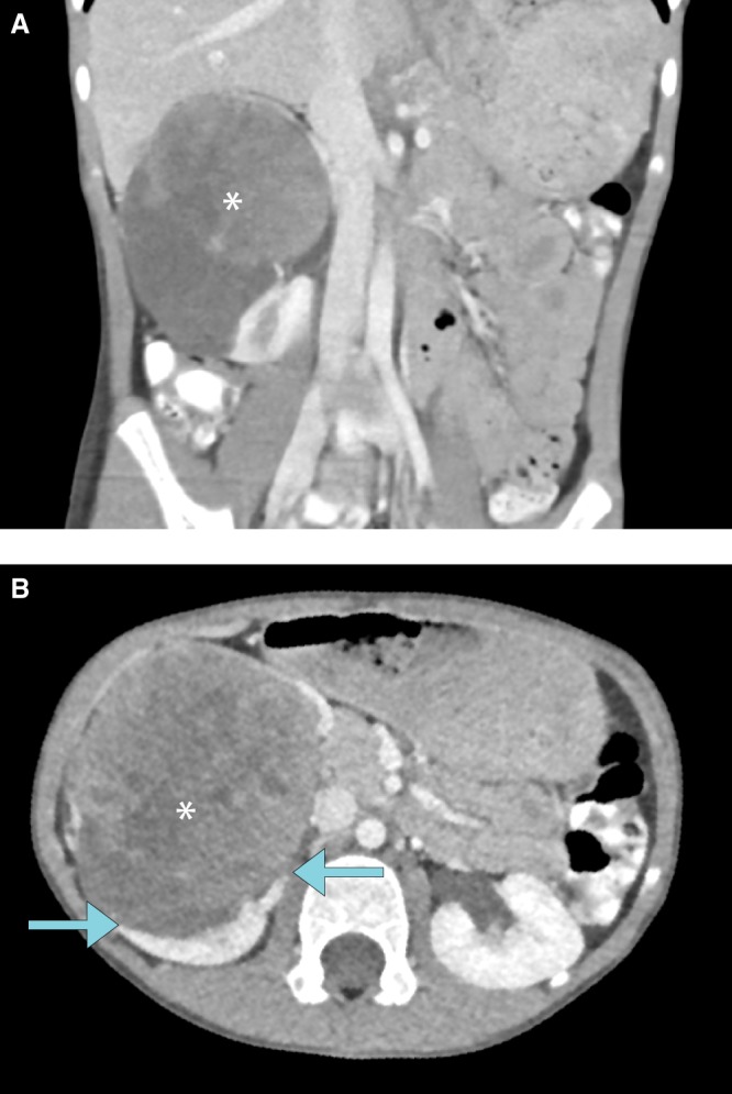
Figure 1.

(A) Coronal contrast-enhanced computed tomography (CECT) image of the abdomen demonstrates a heterogeneously enhancing mass measuring 7.3 × 8.4 × 8.4 cm and arising from the right kidney. Calcific foci are present within the mass (not shown). (B) Origin of the mass from the right kidney, as indicated by the “claw sign” (arrows), is redemonstrated on the axial CT image.
