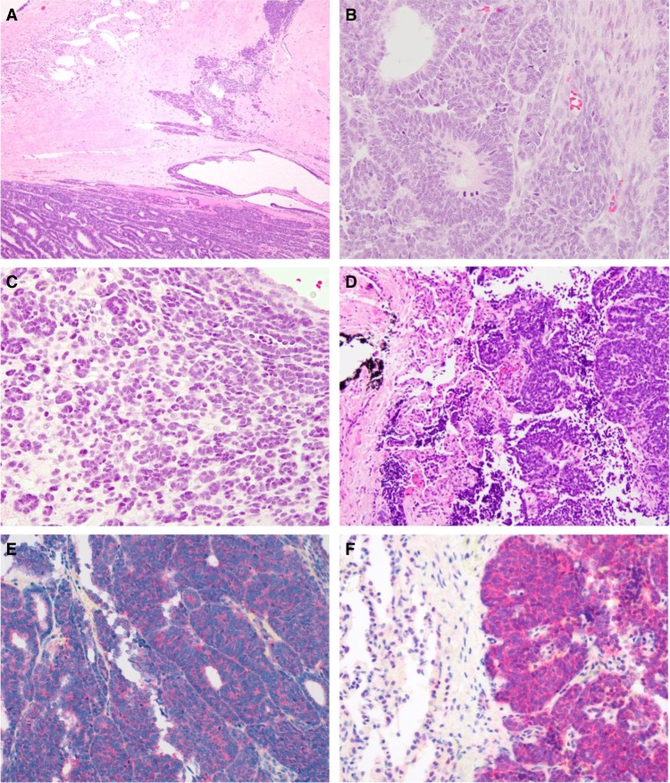
Figure 2.
(A) The nephrectomy revealed an epithelial predominant Wilms tumor (WT). However, there was a differentiated area resembling metanephric adenoma (MA) associated with sclerosis (upper right). Normal kidney adjacent to tumor is at the upper left. (B) WT, triphasic area. Note the mitotic figures. (C) WT, differentiated area mimicking MA. Note the absence of mitotic figures and minimal cytoplasm of the neoplastic cells, which form tubules. (D) Lung metastasis of WT. (E) Cytoplasmic BRAF V600E protein immunoreactivity in the primary renal WT. (F) Cytoplasmic BRAF V600E protein immunoreactivity in the metastatic WT in the lung.

