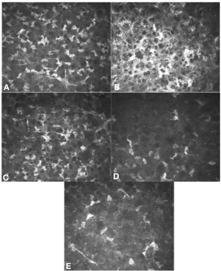
Figure 4.

Laser scanning in vivo confocal microscopy (Heidelberg Retina Tomograph III Rostock Corneal Module (HRTIII) (Heidelberg Engineering, GmBH, Germany)) images of superior anterior corneal stroma, 7mm from the corneal apex. A. Prior to peripheral crosslinking, demonstrating the typical keratocyte appearance and density in a patient with keratoconus.B. One-month post-peripheral crosslinking, demonstrating the expected appearance of a reduction in keratocyte density with hyper-reflective cytoplasm of remaining keratocytes and extracellular lacunae. C. Three months post-peripheral crosslinking, keratocyte repopulation and improvement in hyper-reflectivity of cytoplasm and extracellular lacunae. D. Three months post- Deep Anterior Lamellar Keratoplasty (DALK), early repopulation of keratocytes. E. Twelve months post-DALK, further repopulation of keratocytes
