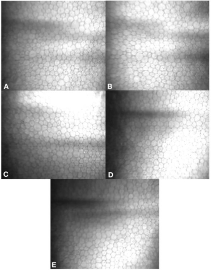
Figure 5.
Slit scanning in vivo confocal microscopy Confoscan 4 (Nidek Technologies, Gamagori, Japan) images of superior corneal endothelium, 7mm from the corneal apex. A. Prior to peripheral crosslinking, mean cell density was 3073 cells/mm2. B. One-month post-peripheral crosslinking, mean cell density was 3028 cells/mm2. C. Three months post-peripheral crosslinking, mean cell density was 3039 cells/mm2. D. Three months post-DALK, mean cell density was 3139 cells/mm2. E. Twelve months post- Deep Anterior Lamellar Keratoplasty (DALK), mean cell density was 3149 cells/mm2

Method for cell count: Only cells within the clearest 200 x 200 µm (333 x 333 pixels) region of an image were counted. Cell counts used the “L” method, whereby cell borders/cell nuclei crossing the left and inferior borders were counted and those crossing the right and superior borders were omitted.
