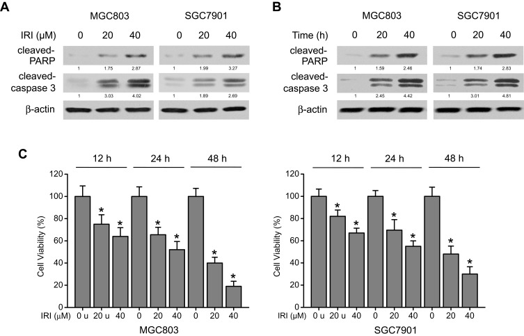
Figure 1.
IRI induces cytotoxicity and apoptosis in gastric cancer cells. (A) MGC803 and SGC7901 cells were treated with IRI (0, 20, or 40 μM) for 24 h or (B) with 20 μM IRI for 0, 12, or 24 h, and cleaved PARP and cleaved caspase 3 protein expression levels were examined by Western blotting. β-actin served as the internal control. (C) MGC803 and SGC7901 cells were incubated with various concentrations of IRI for the indicated periods, and cell viability was determined by MTT assay. *P < 0.05.

