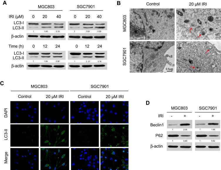
Figure 2.
IRI induces autophagy in gastric cancer cells. (A) MGC803 and SGC7901 cells were treated with IRI (0, 20, or 40 μM) for 24 h, or with 20 μM IRI for 0, 12, or 24 h, and LC3 protein expression was examined by Western blotting. β-actin served as the internal control. (B) TEM detection of autophagosome formation in MGC803 and SGC7901 cells treated with 20 μM IRI for 24 h (red arrows indicate autophagosomes). Scale bar: 0.5 μm. (C) Representative images of LC3-II immunostaining in MGC803 and SGC7901 cells incubated with 20 μM IRI for 24 h. (D) Western blotting analysis of the protein levels of Beclin-1 and P62 in MGC803 and SGC7901 cells incubated with 20 μM IRI for 24 h.

