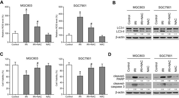
Figure 4.
ROS mediate the IRI-induced autophagy and growth inhibition in gastric cancer cells. (A) MGC803 and SGC7901 cells were pretreated with 5 mM NAC for 1 h and then exposed to 20 μM IRI for 24 h. ROS levels were evaluated with a DCF-DA assay kit, and DCF fluorescence was measured. (B) LC3-I and LC3-II protein amounts were evaluated by Western blotting. β-actin was used as the internal control. (C) The MTT assay was carried out to analyze cell viability. (D) Cleaved-PARP and cleaved-caspase-3 protein expression was examined by Western blotting. β-actin served as the internal control. *P < 0.05 compared to Control group; #P < 0.05 compared to IRI group.

