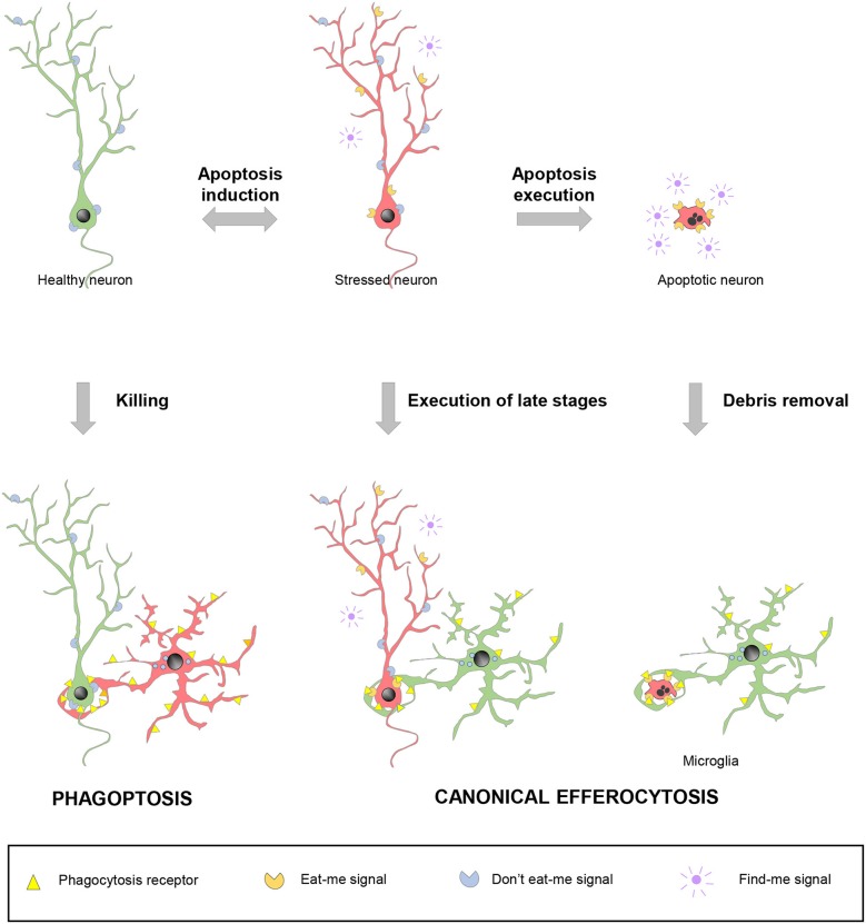
Figure 4.
Spectrum of possibilities between physiological phagocytosis and pathological phagoptosis. Healthy neurons (green) increase their expression of released “find-me” and surface “eat-me” molecules and decrease “don't eat-me” signals under stressful situations. Stressed neurons (red) may revert to the healthy situation or follow up with the completion of the apoptotic program. In non-pathological conditions, apoptotic debris removal rapidly occurs after phagocytosis is executed by professional phagocytes, such as microglia. Under certain circumstances, stressed neurons (red) may be recognized and executed by nearby phagocytes, including microglia, before the apoptotic program has been fully engaged. In contrast to these two cases of canonical efferocytosis, in a third scenario, dysregulation of the “eat-me” and “don't-eat-me” signalization may lead microglia or other phagocytes to directly target for execution healthy, viable neurons in a process termed phagoptosis.

