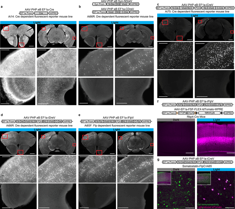
Figure 2. Optogenomic modifications by RecV viruses with spatiotemporal and intersectional cell class specific precision in vivo.
Reporter mice (n=2 per case) received right hemisphere intracerebroventricular (ICV) or retro-orbital (RO) injection of PHP.eB rAAVs followed by light stimulation to the left hemisphere two weeks post injection, and imaging two weeks post light stimulation. (a) Injecting Ai14 mice with AAV-PHP.eB EF1a-Cre virus led to widespread recombination throughout the brain (68928 and 182022 cells per section -CPS). (b) Ai66R mice were ICV injected with a 1:1 mixture of AAV-PHP.eB Syn-NDreV and AAV-PHP.eB EF1a-CDreV (204 and 608 CPS). (c) Ai75 mice were ICV injected with AAV-PHP.eB EF1a-iCreV (1323 and 3649 CPS). (d) Ai66R mice were ICV injected with AAV-PHP.eB EF1a-iDreV (1630 and 2670 CPS). (e) Ai65F mice were injected with AAV-PHP.eB EF1a iFlpV. Scale bars a-e: 1 mm for top images, 200 μm for bottom images (1386 and 2471 CPS). (f) L5 pyramidal neuron-specific Rbp4-Cre mice were injected with a mixture of AAV-PHP.eB EF1a-iFlpV and AAV-PHP.eB EF1a-FSF-FLEX-tdTomato. Scale bar: 250 μm (469 of 478 cells in L5). (g) Somatostatin FlpO mouse line, Sst-IRES-FlpO, crossed with a Cre/Flp double-dependent fluorescent reporter mouse line, Ai65, was RO injected with AAV-PHP.eB EF1a-iCreV and light was delivered to the left hemisphere. Specific intersectional light-induced recombination was observed in somatostatin-positive inhibitory interneurons as revealed by immunohistochemistry (100% reporter positive cells (119) were Sst positive (313); 38.1% Sst cells were reporter positive.). Scale bar: 250 μm for inset and 75 μm for zoomed images. All in vivo light activation was applied through the skull on the left hemisphere (opposite ICV injection sites in those cases). For a-e, two coronal planes are shown for each injection (top row) with enlarged views (lower two rows) for areas indicated by red boxes.

