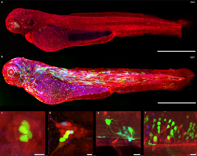
Figure 3. CreV allows tight optogenomic modifications in multiple tissues of Danio rerio.
(a) Confocal images of dark reared Zebrabow zebrafish larva (3dpf) co-injected with N-CreV and C-CreV plasmids (see methods section for details). No recombination was observed. In the default state, RFP is expressed in all cells. Apparent green signal is due to autofluorescence. (b) Confocal images of a Zebrabow larva co-injected with N-CreV and C-CreV plasmids and immediately exposed to light. CreV-mediated recombination in multiple tissue types is reflected as expression of YFP and CFP: (c) lateral line hair cells, (d) trigeminal ganglion, (e) spinal neuron and glial cells, and (f) hindbrain neurons. Scale bars: (a-b) 500μm, (c-f) 10μm.

