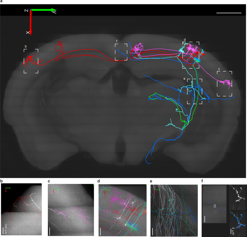
Figure 4. Cortical pyramidal cells (PCs) labeled with CreV and reconstructed at the whole-brain level.
(a) Eight reconstructed PCs in a mouse somatosensory cortex include three layer 2/3 PCs (in pink) with ipsilateral cortico-cortical projections, two layer 2/3 PCs (in red) with contralateral cortico-cortical projections, and three L5 thick tufted PCs (one green, one blue, one light blue) with ipsilateral cortico-subcortical projections. Local axonal clusters are incomplete because labeling at the somata region is still too dense in this brain for tracing fine axonal branches. The eight reconstructed PCs are superimposed onto a coronal brain plane located 5201–5400 μm posterior to the olfactory bulb (scale bar: 1 mm). (b-f) Enlarged views of areas outlined by dashed boxes in a, with reconstructions (in colors) superimposed on original images with GFP fluorescence shown as white. In f, the two panels on the right (without reconstruction in white, with reconstruction in blue) are enlarged views of the boxed area in the left panel, showing the high-resolution details of a segment of axon with enlarged boutons clearly visible. The whole brain image stack is composed of 12089 images, resolution of XYZ, 0.3 × 0.3 × 1 μm.

