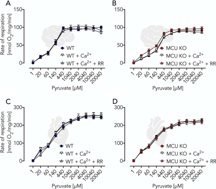
Figure 2.
Validation of Ca2+ independence of pyruvate-induced respiratory activation using isolated brain and heart mitochondria. A and B, respiratory rates of WT and MCU KO brain mitochondria (0.06 mg/ml) incubated in EGTA buffer supplemented with ADP (2 mm) and malate (2 mm) and additions of pyruvate, Ca2+ (800 nm), and RR as indicated. C and D, respiratory rates of WT and MCU KO heart mitochondria (0.04 mg/ml) under conditions as described for A and B. All data are shown as mean ± S.E. (error bars) of n = 5 experiments.

