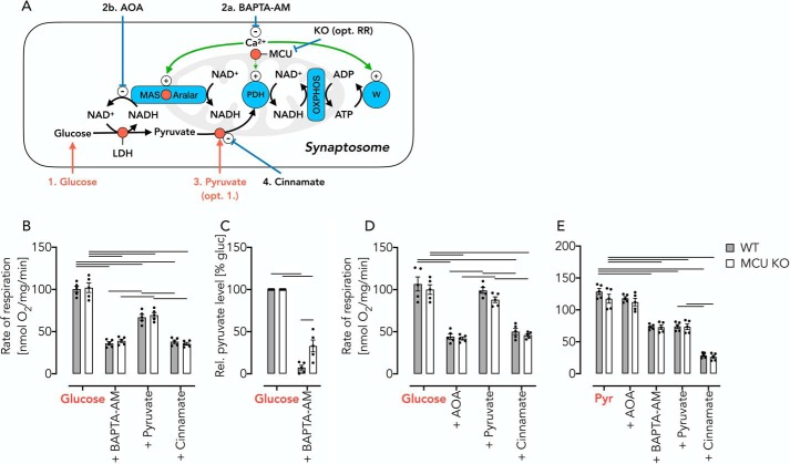
Figure 6.
Detection of reversible pyruvate starvation after AOA– or BAPTA-AM–induced inhibition of the endogenous MAS in intact synaptosomes. A, schematic of experimental set-up. The numbers indicate the order of addition. B, respiratory rates of WT and MCU KO synaptosomes in KRB supplemented with glucose/lactate, 20 min after the addition of BAPTA-AM (50 μm), after the addition of pyruvate (10 mm), and after inhibition of the mitochondrial pyruvate carrier using Cin. Of note, BAPTA-AM–induced Ca2+ depletion affects both the cellular workload and the substrate supply for OXPHOS. C, pyruvate levels in incubations of synaptosomes with glucose (10 mm)/lactate (10 mm) before and after the addition of BAPTA-AM (50 μm). D, respiratory rates in WT and MCU KO synaptosomes using AOA instead of BAPTA-AM to induce a state of pyruvate starvation. E, respiratory rates in WT and MCU KO synaptosomes measured in KRB supplemented with pyruvate (Pyr) under the conditions indicated. Here, BAPTA-AM–induced Ca2+-depletion affects workload but not mitochondrial substrate supply. All data are shown as mean ± S.E. (error bars) of n = 5 experiments. Horizontal bars in B, D, and E, p < 0.05 for comparison as indicated, determined using two-way ANOVA with Tukey's multiple-comparison test. Horizontal bars in C, p < 0.05 for comparison as indicated, determined using two-way ANOVA with Sidak's multiple-comparison post hoc test.

