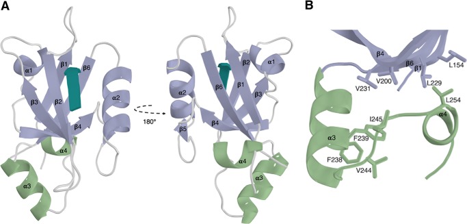
Figure 1.
The extended PDZ2 domain of NHERF1 in complex with the CFTR PDZ-binding motif. A, the extended helical regions (green) couple to the canonical PDZ domain (purple) at a region distal to the PDZ-binding motif (teal). B, hydrophobic interactions formed between the α3 and α4 and β1, β4, and β6 from the core PDZ-fold.

