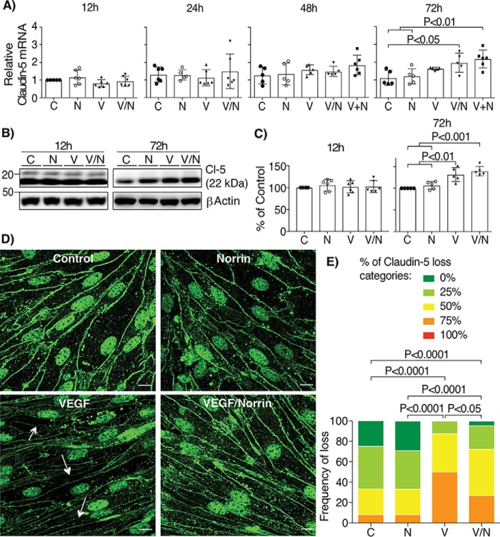
Figure 8.
Norrin and VEGF both increase the expression of claudin-5, but only norrin promotes its localization at cell contacts. BREC stimulated with vehicle (control, C), norrin (N), VEGF (V), both (V/N), or norrin after VEGF (V+N) for 12, 24, 48, and 72 h were processed for qRT-PCR with specific primers for claudin-5; n = 5–6 (A). All samples were normalized to control (C) for 12 h. B and C, Western blotting of claudin-5 (Cl-5); n = 5–6. D, immunostaining of claudin-5 in BREC stimulated at 72 h; n = 3. Arrows point to typical sites of tight junction disruption; scale bar, 10 μm. E, quantification of four images per experiment by masked scoring combined from three individuals. Data show the frequency of claudin-5 loss at the cell contacts ranked in five categories, where 100% corresponds to completely lost. p values were calculated by one-way ANOVA, followed by Tukey's post hoc test. Error bars, S.D.

