. 2020 Mar 2;9:e51065. doi: 10.7554/eLife.51065

Figure 2. The degradation of MAD QC substrates requires the ubiquitin-proteasome system.

(A, B) CHX chase for the indicated times at 37°C assessing the turnover of sam35-2HAts or sen2-1HAts (pMM157 or 160, respectively) in WT (WCG4a) and pre1-1 pre2-2 proteasome mutant (WCG4-11/21a) cells (A) or WT (CIM) and cim3-1 proteasome mutant cells (B). Proteins were detected by immunoblotting. Graphed below is the mean and SD of the PGK-normalized HA signal at each time point for three biological replicates. (C) Lysates from the strains used in A were fractionated at 12,000xg into mitochondrial pellets (P) and post-mitochondrial supernatants (S) after incubation at 37°C for the indicated times. Fractions were subject to immunoblotting with antibodies to HA, PGK, and PORIN. (D) CHX chase for the indicated times at 37°C assessing the turnover of sam35-2HAts or sen2-1HAts (pMM157 or 160, respectively) in a uba1-204 strain relative to its isogenic WT strain. (E) Ubiquitination of sam35-2HAts and sen2-1HAts was assessed by immunoprecipitation (IP) from lysates of the strains used in A with anti-HA agarose, followed by immunoblotting with ubiquitin antibodies. 1% of IP input lysate was reserved and also analyzed by immunoblotting. (F) Ubiquitination of sam35-2HAts and sen2-1HAts was assessed by IP from lysates of the strains used in A using tandem ubiquitin-binding entities (TUBE) agarose, followed by immunoblotting with HA antibody. 2.5% of the TUBE input lysate was reserved and analyzed by immunoblotting.

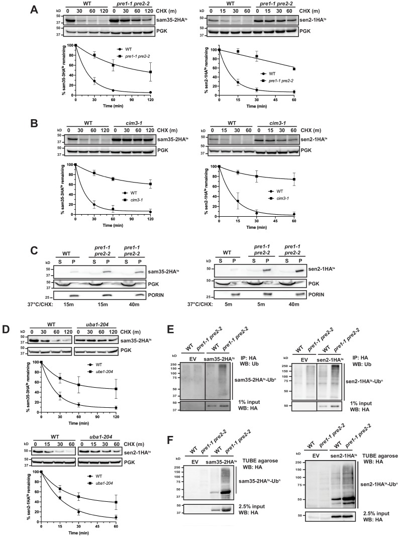
Figure 2—source data 1. Quantifications of cycloheximide chases.

Figure 2.

Figure 2—figure supplement 1. The degradation of MAD QC substrates requires the ubiquitin-proteasome system.

Figure 2—figure supplement 1.

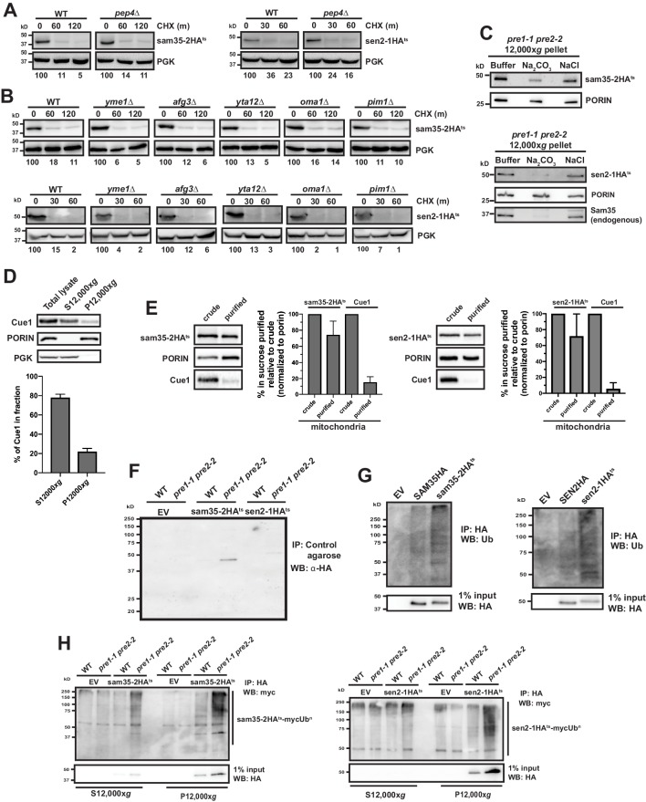
(A, B) CHX chase for the indicated times at 37°C assessing the turnover of sam35-2HAts or sen2-1HAts (pMM157 or 160, respectively) in WT (BY4741) and pep4Δ (A) or yme1Δ, afg3Δ, yta12Δ, oma1Δ, or pim1Δ (B) cells. Proteins were detected by immunoblotting. Percentage of each ts- substrate remaining is labeled for each time point under blots. (C) Sodium carbonate (Na2CO3) extraction of sam35-2HAts or sen2-1HAts (pMM157 and 160, respectively) from the 12,000xg mitochondrial pellet of pre1-1 pre2-2 (WCG4-11/21a) cells. Control treatments of sodium chloride (NaCl) and buffer are shown for comparison. Proteins were detected with immunoblotting for HA, PORIN, and Sam35 (in analysis of sen2-1HAts only). (D) Total lysate from pre1-1 pre2-2 (WCG4-11/21a) cells expressing sam35-2HAts (pMM157) were fractionated at 37°C into mitochondrial pellets (P12,000xg) and post-mitochondrial supernatants (S12,000xg). Fractions were subject to immunoblotting with antibodies to Cue1, PGK, and PORIN. The Cue1 signal in each fraction was quantified and the mean plus standard deviation (SD) of the percentage of the total Cue1 signal is graphed below (N = 3). (E) P12,000xg mitochondrial fractions (‘crude’) isolated from pre1-1 pre2-2 (WCG4-11/21a) cells expressing sam35-2HAts or sen2-1HAts (pMM157 and 160, respectively) were further purified by sucrose gradient fractionation (‘purified’). The HA (sam35-2HAts or sen2-1HAts) and Cue1 signals in crude and purified mitochondrial fractions were quantified and normalized to the PORIN signal. The crude mitochondrial signal was set to 100% and the mean plus SD of the percentage in purified mitochondria relative to crude is graphed (N = 3). (F) Negative control experiment for Figure 2E. Cell lysates from pre1-1 pre2-2 (WCG4-11/21a) cells expressing sam35-2HAts, sen2-1HAts, or empty vector (EV; pMM157, pMM160, or pRS315, respectively) were precipitated with unconjugated TUBE control agarose (containing no ubiquitin-binding domains), followed by immunoblotting with HA antibody. (G) Ubiquitination was assessed by IP with anti-HA agarose from lysates of WT (BY4741) cells expressing empty vector (EV; pRS315), SEN2HA (pMM159), sen2-1HAts (pMM160), SAM35HA (pMM158), or sam35-2HAts (pMM157), followed by immunoblotting with ubiquitin antibody. 1% of the IP was reserved and analyzed by immunoblotting for the unmodified proteins. (H) Ubiquitination in the 12,000xg mitochondrial pellet (P12,000xg) or post-mitochondrial supernatant (S12,000xg) was assessed by IP from each fraction with anti-HA agarose from WT (WCG4a) and pre1-1 pre2-2 (WCG4-11/21a) cells expressing myc-Ub (pSM3666) and either empty vector (EV; pRS315), sam35-2HAts (pMM157), or sen2-1HA (pMM160), followed by immunoblotting with c-myc antibody. 1% of IP input lysate was reserved and analyzed by immunoblotting.