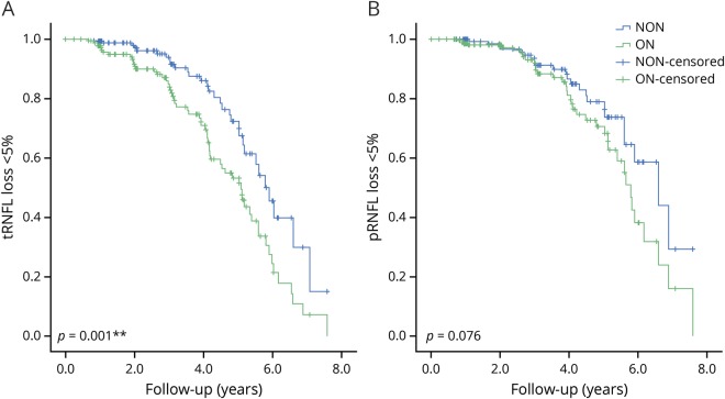
Figure 1. ON eyes showed a faster rate of RNFL loss in the temporal disc area.
Comparisons of progressive tRNFL (A) and pRNFL (B) thinning between ON and fellow NON eyes in patients with unilateral ON (n = 51) using Kaplan-Meier curves and the log-rank test. More than 5% of RNFL thickness loss as compared to the baseline was defined as the end point. NON = non-ON; ON = optic neuritis; pRNFL = global peripapillary retinal nerve fiber layer; RNFL = retinal nerve fiber layer; tRNFL = temporal RNFL.

