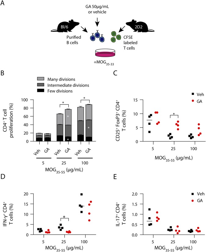
Figure 5. GA-treated B cells preferentially generate T regs, whereas development of proinflammatory T cells is diminished.
(A) Naive B cells purified from WT mice were incubated with 50 μg/mL GA or vehicle at 37°C for 3 hours. After washing, B cells were cocultured with CFSE-labeled myelin-specific (2D2) naive T cells in the presence of 5, 25, or 100 μg/mL MOG peptide35-55. (B) T-cell proliferation was evaluated by CFSE dilution and stratified by division frequency as follows: few divisions (1–2; black), intermediate divisions (3; medium gray), and many divisions (≥4; light gray). T-cell divisions are shown as mean ± SEM; n = 4; *p < 0.05; Mann-Whitney U test. Differentiation of myelin-specific naive T cells into (C) Treg cells (CD25+FoxP3+CD4+) or (D) Th1- (IFN-γ+CD4+) and (E) Th17 cells (IL-17+CD4+) was analyzed by FACS (data given as median; n = 4; *p < 0.05; Mann-Whitney U test). GA = glatiramer acetate.

