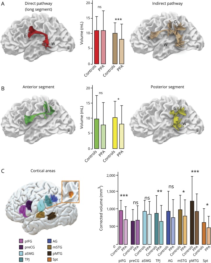
Figure 2. Group differences between patients with primary progressive aphasia (PPA) and controls in the perisylvian white matter and cortical areas.
(A–C) Error bars indicate 2 SDs from the mean. *Independent t test significance level p < 0.05. **Independent t test significance level p < 0.01. ***Independent t test significance level p < 0.001. AG = angular gyrus; aSMG = anterior supramarginal gyrus; B = Broca territory; G = Geschwind territory; mSTG = middle part of the superior temporal gyrus; ns = not significant; pIFG = posterior inferior frontal gyrus; pMTG = posterior middle temporal gyrus; preCG = precentral gyrus; Spt = Sylvian parietal temporal area; TPJ = temporo-parietal junction; W = Wernicke territory.

