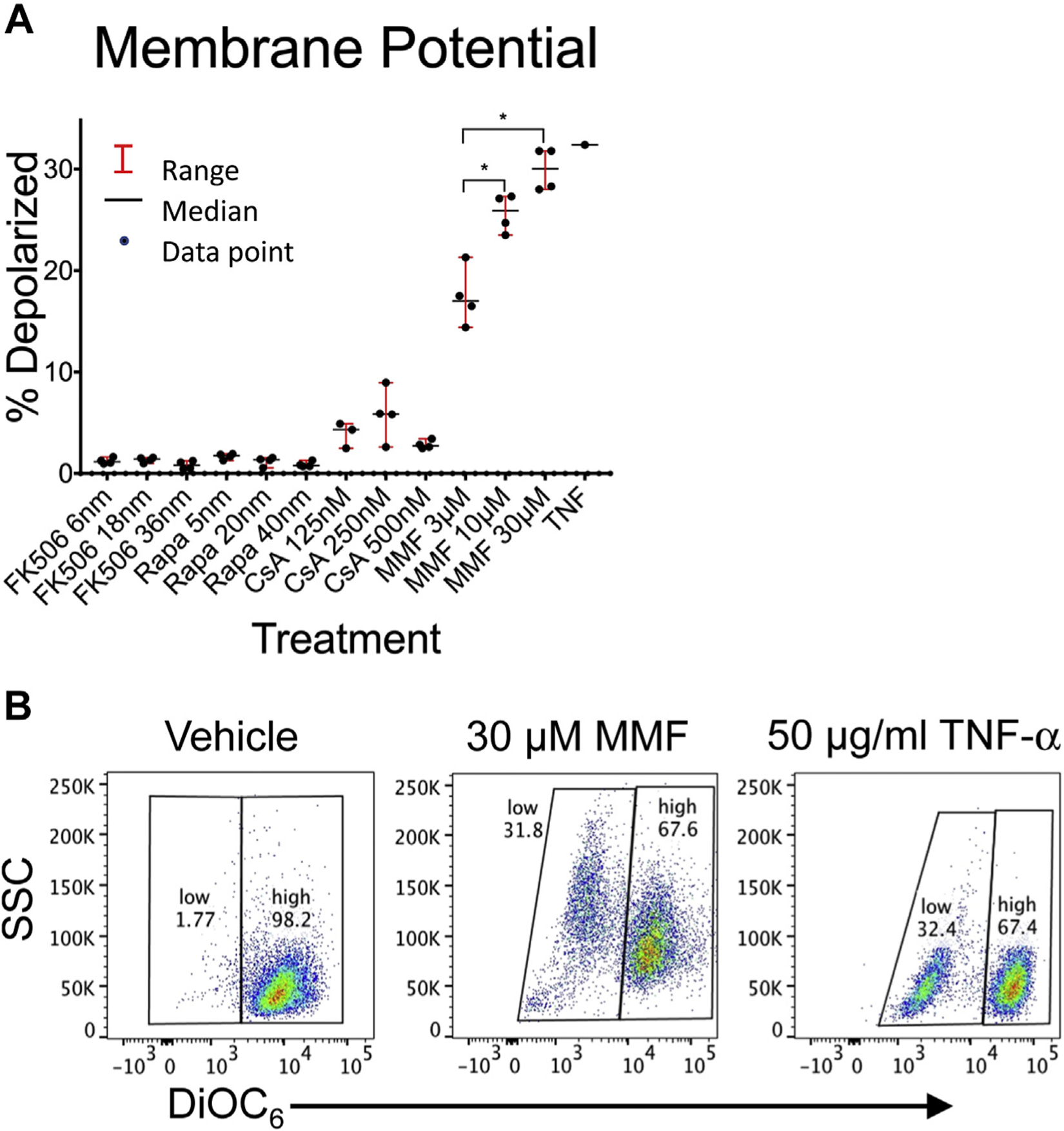
Fig. 1 –

Effect of immunosuppression reagents on T-cell mitochondrial membrane potential. Jurkat cells were incubated for 24 h with therapeutic and supratherapeutic levels of tacrolimus (FK506), rapamycin (Rapa), cyclosporine A (CsA), and mycophenolate mofetil (MMF). After 24-h incubation, cells were stained with DiOC6 and analyzed by flow cytometry for mitochondrial membrane depolarization as assessed by decreased DiOC6 fluorescence. (A) The percentage of cells with reduced DiOC6 staining is shown for each drug and concentration in quadruplicate. The median and range of each data set is also shown. (B) Representative flow cytometry after incubation with vehicle, 30 μM MMF, and TNF-α control. Comparisons between treatment and vehicle control groups and between each treatment groups were performed using a two-tailed unpaired Student’s t-test. *P-values were adjusted for multiple comparisons using the Bonferroni correction with a standard significant P value of 0.05. Significant P-values after correction are marked with an asterisk. TNF-α = tumor necrosis factor alpha.
