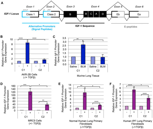
Figure 5. TGFβ upregulates the Class-1 IGF-1 promoter in fibroblasts and pulmonary fibrosis tissue.
(A) A schematic diagram of IGF-1 gene locus [93, 94]. (B) RT-qPCR of IGF-1 gene expression via Class-1 (C1) and Class-2 (C2) alternative promoters following 12 hours (peak IGF-1 induction by TGFβ) treatment with TGFβ (+) or vehicle (−) in AKR-2B (n=3) murine fibroblasts. (C) RT-qPCR gene expression analysis of IGF-1’s C1 and C2 promoters in whole lung tissue from intratracheal Bleomycin (BLM, n=5) treated mice compared to Saline (n=5). (D-F) IGF-1 promoter usage following 24 hours (peak time of TGFβ-dependent stimulation of IGF-1 in human fibroblasts) treatment of (D) MRC5 (n=3), (E) Normal Human Lung Primary Fibroblasts (NHLF, n=5), and (F) Human Idiopathic Pulmonary Fibrosis Primary Fibroblasts (IPF, n=4). Data are presented as means −/+ Standard Error of the Mean (SEM) for the number of biological replicates indicated (n). Statistical significance was determined after computing single factor ANOVA and/or unpaired two-tailed Student’s t-test (p<0.05 (*), p<0.01 (**), p<0.005 (***), p<0.001 (****). Results demonstrate that of the two alternative promoters required for transcription initiation of IGF-1, TGFβ increases transcription of the C1 promoter significantly more than the C2 in both murine and human myofibroblasts.

