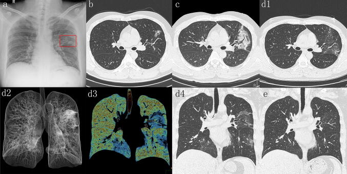
Fig. 1.
a CXR shows infiltrate in the left middle lung field (red square). b High resolution CT with 1 mm thickness after admission at disease onset shows patchy ground-glass opacity in left upper lobe. c Follow-up CT 3 days after admission shows evolution to a segmental mixed pattern of ground-glass opacities and consolidation that grow larger with air bronchogram in left upper lobe. d Follow-up CT 7 days after admission (d1, axial image; d2, ray-summation image; d3, pseudo color MIP; d4, coronal image) shows multifocal bilateral ground-glass opacities and improvement of mixed ground-glass opacities and consolidation in left upper lobe. e Coronal MPR image obtained 2 weeks after admission shows marked improvement of multifocal ground-glass opacities in both lungs

