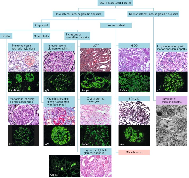
Fig. 2. Categorization of MGRS-associated renal lesions.
Monoclonal gammopathy of renal significance (MGRS)-associated renal lesions (blue boxes) are initially separated by the presence or absence of monoclonal immunoglobulin deposits in kidney biopsy samples. They are further subcategorized by the ultrastructural characteristics of the deposits into organized and non-organized. Organized deposits are further subdivided into fibrillar, microtubular and inclusions or crystalline categories. Images of typical histological sections stained with haematoxylin and eosin (H&E), periodic acid–Schiff or Masson trichrome stain and Congo red (top) are paired with immunofluorescence studies of frozen tissue sections (bottom) to reveal the specific immunoglobulin species. Pink box: the miscellaneous category represents polyclonal glomerulopathies that sometimes present with monoclonal immunoglobulin deposits, such as monotypic membranous nephropathy and monotypic anti-glomerular basement membrane disease. Purple box: thrombotic microangiopathy currently has a provisional status as an MGRS-associated lesion pending further evidence. Because this lesion has no immunoglobulin deposits and is best identified by electron microscopy, the immunofluorescence and H&E stained sections were replaced by an electron micrograph. LCPT, light-chain proximal tubulopathy; MIDD, monoclonal immunoglobulin deposition disease; PGNMID, proliferative glomerulonephritis and monoclonal immunoglobulin deposits.

