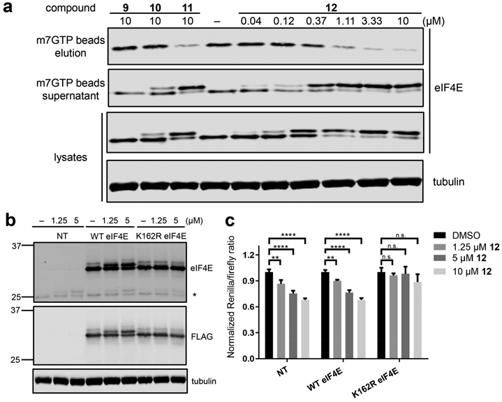
Figure 3.

Covalent inactivation of eIF4E in cells. (a) Jurkat cells were treated with 9–12. After 3 h, cell lysates were prepared, and an aliquot of each sample was analyzed by Western blotting for eIF4E and tubulin (“lysates”). Remaining lysate samples were enriched with m7GTP-agarose beads, and the bound (“elution”) and unbound (“supernatant”) fractions were analyzed by Western blotting. (b) HEK293T cells stably overexpressing WT or K162R FLAG-eIF4E or nontransduced cells (NT) were treated with 12 for 30 min. Cell lysates were prepared and analyzed by Western blotting (* denotes endogenous eIF4E). (c) Stable cell lines from (b) were treated with 12 prior to transfection with a bicistronic plasmid comprising a cap-dependent cistron (Renilla luciferase) followed by a cap-independent cistron (firefly luciferase). Cells were incubated with 12 or DMSO for 6 h, after which the Renilla and firefly luciferase activities were measured (Figure S9). Data are means ± SEM (n = 3). **, P < 0.01; ****, P < 0.0001.
